
Inferior pharyngeal constrictor full#
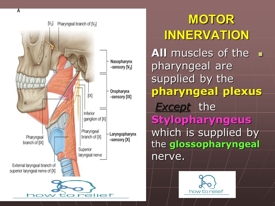
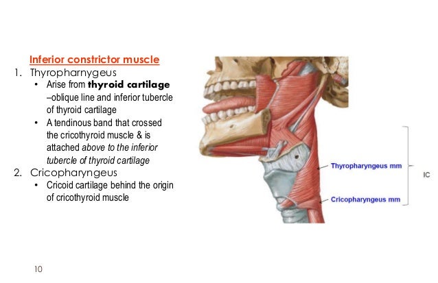
Although the UES is often described as a 2- to 4-cm zone of high pressure encompassing the IPC, CP, and the upper esophagus, in practice, the UES and CP are interchangeable (see Chapter 5 for full description of the UES). This muscle forms the majority of the upper esophageal sphincter (UES). Its most caudal portion is commonly referred to as the cricopharyngeus (CP) muscle. The inferior pharyngeal constrictor (IPC) is the thickest and best developed of the three pharyngeal constrictors. Chhetri MD, in Dysphagia Evaluation and Management in Otolaryngology, 2019 Introduction Some of the muscles also help with speaking.Andrew Vahabzadeh-Hagh MD, Dinesh K. They all act on the pharynx, either constricting or elevating it.Īll the muscles of the pharynx help to push the food bolus towards the esophagus during the act of swallowing either by contracting, or by shortening the pharynx itself. They are all innervated by the pharyngeal plexus and pharyngeal branch of the vagus nerve, except the stylopharyngeus which is innervated by the glossopharyngeal nerve. Insertions: Blends with pharyngeal constrictors, lateral glossoepiglottic fold, posterior border of thyroid cartilage Origins: Medial base of styloid process of temporal bone Insertions: Blends with palatopharyngeus muscle Origins: Inferior/cartilaginous part of auditory (Eustachian) tube

Insertions: Posterior border of thyroid cartilage, blends with contralateral palatopharyngeus muscle Origins: Posterior border of hard palate, palatine aponeurosis Insertions: Median pharyngeal raphe (Thyropharyngeal part), Blends inferiorly with circular esophageal fibres (Cricopharyngeal part) Origins: Oblique line of thyroid cartilage (Thyropharyngeal part), Cricoid cartilage (Cricopharyngeal part) Insertions: Median pharyngeal raphe, Blends with superior and inferior pharyngeal constrictors Origins: Stylohyoid ligament, Greater and lesser horn of hyoid bone Insertions: Pharyngeal tubercle on basilar part of occipital bone Origins: Pterygoid hamulus, pterygomandibular raphe, posterior end of mylohyoid line of mandible Key facts about the muscles of the pharynx Superior pharyngeal constrictor There are six pharynx muscles in total that can be divided into two groups: Pharyngeal constrictors and longitudinal muscles Attachments Laryngopharynx - posterior to the larynx.Oropharynx - posterior to the oral cavity.Nasopharynx - posterior to the nasal cavity.

Notice how the nasopharynx connects the oral and nasal cavities, allowing a person to breathe through the nose.īased on its anterior relations, the pharynx consists of three regions: Pharyngeal plexus: receives branches of vagus nerve (CN X), glossopharyngeal nerve (CN IX) and maxillary nerve (CN V2) Longitudinal muscles: Palatopharyngeus, salpingopharyngeus, stylopharyngeusįacial artery, lingual artery, maxillary artery (branches of external carotid artery) Pharyngeal constrictors: Superior, middle and inferior muscles In this page, we will learn more about the anatomy of the pharynx and its functions, including its main regions and muscles. In addition, they also help with swallowing and speaking. The functions of the pharynx are accomplished by two sets of muscles which help push the food bolus further down the digestive tract. Essentially, it forms a continuous muscular passage for air, food, and liquids to travel down from your nose and mouth to your lungs and stomach. The pharynx, more commonly known as the throat, is a 12-14 cm, or 5 inch, long tube extending behind the nasal and oral cavities until the voice box ( larynx) and the esophagus.


 0 kommentar(er)
